Runner's Knee
Patellofemoral pain syndrome, commonly known as runner’s knee, is one of the most frequently encountered musculoskeletal complaints in active individuals. It is estimated to account for roughly 25% of all running-related injuries, and is also common among cyclists, hikers and people who spend extended time climbing stairs or squatting1. The condition involves pain arising from the patellofemoral joint, which is the articulation between the kneecap (patella) and the groove at the front of the thigh bone (femur).
Causes of Runner’s Knee
Runner’s knee develops when the patella does not track smoothly within the femoral groove during movement. Rather than gliding centrally, the kneecap may shift laterally or tilt, producing excessive pressure and friction on the cartilage surfaces of the joint. Several factors contribute to this abnormal tracking:
- Muscle imbalances: weakness of the hip abductors, hip external rotators and the VMO (vastus medialis oblique, the inner quad muscle) allows the femur to rotate inward, pulling the patella off its ideal path
- Training errors: rapid increases in mileage or intensity, hill running and high-volume stair work place repetitive compressive load on the joint before tissues have adapted
- Biomechanical factors: excessive foot pronation, genu valgum (knock-knee alignment) and a wide Q-angle can all alter patellar tracking
- Tight lateral structures: a shortened iliotibial band or lateral retinaculum pulls the kneecap outward, increasing lateral facet pressure
In chronic cases, sustained abnormal loading can cause breakdown of the articular cartilage lining the back of the patella, transitioning the condition from irritation to structural degeneration.
Risk Factors
- Running, cycling or other repetitive knee-flexion activities
- Sudden increase in training volume or intensity
- Female sex (wider pelvis increases Q-angle)
- Young and adolescent athletes with rapid growth
- Flat feet or excessive pronation
- Previous knee injury or surgery
- Prolonged kneeling or squatting at work
Symptoms
The hallmark symptom is a diffuse, aching pain around or behind the kneecap. Pain is typically provoked or worsened by:
- Running, especially downhill
- Descending stairs or inclines
- Prolonged sitting with the knees bent (the “theatre sign”)
- Squatting and lunging movements
Mild swelling around the kneecap may be present. A grinding or clicking sensation (crepitus) is sometimes felt with knee flexion and extension. Unlike a ligament or meniscal injury, sharp instability is not usually a feature.
Diagnosis
Diagnosis is primarily clinical, based on a thorough history and physical examination. Assessment includes evaluation of patellar tracking, hip and quadriceps strength, lower-limb alignment and flexibility. Provocative tests such as the Clarke’s sign and patellar compression test help confirm patellar involvement. Imaging is not routinely required, but an X-ray may be ordered to rule out bony abnormalities, and an MRI can assess the degree of cartilage damage or identify other intra-articular pathology in cases that do not respond as expected to initial treatment2.
Conventional Treatment
First-line management centres on relative rest from aggravating activities, ice application and physiotherapy. A structured rehabilitation program addressing hip abductor and VMO strengthening, flexibility work for the hip flexors and IT band, and gait retraining forms the backbone of conservative care. Patellar taping techniques (such as McConnell taping) and braces with a patellar cut-out can offload the joint during the recovery period. Custom or prefabricated orthotics may help if pronation is a contributing factor.
Oral non-steroidal anti-inflammatory drugs (NSAIDs) are frequently used for short-term pain relief. While useful acutely, NSAIDs do not address the underlying biomechanical drivers of the condition and carry gastrointestinal and cardiovascular risks with prolonged use. Corticosteroid injections into the knee joint may reduce inflammation temporarily, but repeated injections are associated with cartilage degradation and are not recommended as a long-term strategy3. For the subset of patients with chronic, recalcitrant symptoms who have not improved with conservative care, regenerative treatment approaches offer a meaningful alternative.
Regenerative Treatment Options
When runner’s knee becomes a persistent, chronic problem despite physiotherapy and rest, regenerative injection therapies can support tissue healing and provide lasting pain relief. These treatments aim to address the underlying structural and cellular dysfunction rather than simply suppressing symptoms.
Prolotherapy involves the injection of a concentrated dextrose (sugar) solution into the affected joint and surrounding ligamentous or tendinous attachments. The solution triggers a controlled local healing response, promoting the repair of weakened connective tissue and restoring mechanical stability to the patellofemoral joint. Read more about prolotherapy and regenerative injection therapy.
Prolozone therapy combines ozone gas with proliferative nutrients and is injected into the joint and surrounding soft tissues. Ozone is a potent stimulator of tissue oxygenation and antioxidant defences, and has been shown to reduce pain and improve joint function in chronic musculoskeletal conditions. Read more about prolozone therapy.
Platelet-rich plasma (PRP) therapy involves drawing a small sample of a patient’s blood, concentrating the platelet-rich fraction by centrifugation and injecting it at the site of injury. Platelets carry a rich array of growth factors that directly stimulate cartilage repair and soft-tissue regeneration. Research supports the use of PRP for knee pain associated with cartilage pathology, with studies demonstrating improvements in pain and function that are sustained over time4. Read more about PRP therapy.
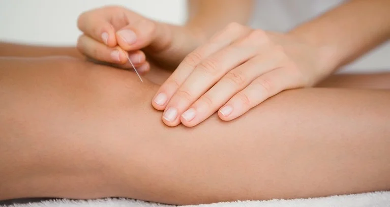
Acupuncture is a well-established complementary treatment for musculoskeletal pain. By stimulating local tissue blood flow and modulating pain signalling pathways, acupuncture can serve as an effective adjunct to rehabilitation, helping to reduce pain and improve function during the recovery process.
To learn more about how these treatments are applied to knee and sports-related injuries, visit the sports medicine page.
References
- Petersen W, Ellermann A, Gösele-Koppenburg A, et al. Patellofemoral pain syndrome. Knee Surg Sports Traumatol Arthrosc. 2014;22(10):2264-2274.
- Crossley KM, Stefanik JJ, Selfe J, et al. 2016 Patellofemoral pain consensus statement from the 4th International Patellofemoral Pain Research Retreat. Br J Sports Med. 2016;50(14):839-843.
- Warden SJ, Metcalf BR, Kiss ZS, et al. Low-intensity pulsed ultrasound for chronic patellar tendinopathy: a randomized, double-blind, placebo-controlled trial. Rheumatology (Oxford). 2008;47(4):467-471.
- Kon E, Buda R, Filardo G, et al. Platelet-rich plasma: intra-articular knee injections produced favorable results on degenerative cartilage lesions. Knee Surg Sports Traumatol Arthrosc. 2010;18(4):472-479.