Sacroiliac Joint Dysfunction
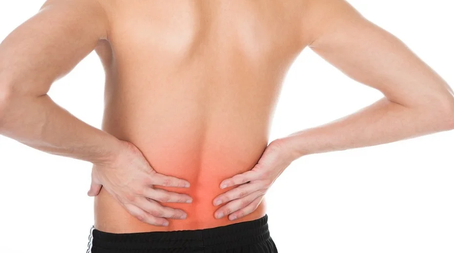
Sacroiliac (SI) joint dysfunction is one of the most frequently overlooked causes of low back and buttock pain, estimated to account for 15 to 30% of chronic low back pain cases.1 The sacroiliac joints connect the sacrum (the triangular bone at the base of the spine) to the ilium bones of the pelvis on each side. These joints are held together primarily by strong ligaments and bear the significant task of transferring load between the spine and the lower limbs. When the ligaments supporting the SI joint become lax or damaged, or when the joint itself becomes inflamed or degenerated, pain and instability can result. Because SI joint pain can mimic disc herniation, hip pathology and other causes of low back pain, it is commonly missed in conventional diagnostic workups.
Causes of SI Joint Dysfunction
The SI joint can become painful through several different mechanisms:
- Ligament laxity: The ligaments surrounding the SI joint are its primary stabilizers. When these ligaments are stretched or injured, excessive or abnormal movement of the joint can cause pain and inflammation.
- Pregnancy and postpartum changes: Hormonal changes during pregnancy increase ligament laxity throughout the pelvis, which can destabilize the SI joint. This effect may persist after delivery.
- Trauma: A fall onto the buttocks, a motor vehicle accident or a misstep can directly injure the SI joint ligaments.
- Leg length discrepancy: A difference in leg length alters gait mechanics and places asymmetric stress on the SI joints over time.
- Degenerative arthritis: Like other joints, the SI joint is susceptible to osteoarthritis, which can cause joint surface deterioration and chronic pain.
- Inflammatory arthritis: Conditions such as ankylosing spondylitis can specifically target the sacroiliac joints.
Risk Factors
Certain factors increase the likelihood of developing SI joint dysfunction:
- Female sex (women are at higher risk due to wider pelvic anatomy and hormonal influences on ligament laxity)
- Pregnancy and the postpartum period
- History of pelvic or low back trauma
- Previous lumbar spine surgery, which can alter load distribution to the SI joints
- Occupations or activities involving repetitive single-leg loading or asymmetric movement
- Leg length discrepancy
- Hypermobility syndromes
Symptoms of SI Joint Dysfunction
SI joint pain is characteristically one-sided, though bilateral cases occur. Common symptoms include:
- Pain in the low back, buttock or upper posterior thigh on the affected side
- Pain that worsens with prolonged sitting or standing
- Difficulty with transitions such as rising from a chair or rolling over in bed
- Pain when climbing stairs or walking on uneven ground
- Aching or sharp pain with single-leg weight-bearing activities
- Pain that may radiate into the groin or down the posterior thigh, but typically not below the knee
Unlike lumbar disc herniation, SI joint dysfunction rarely produces neurological symptoms such as numbness, tingling or significant leg weakness below the knee.
Diagnosis
There is no single definitive test for SI joint dysfunction, which contributes to how often it is missed. Diagnosis is based on a combination of clinical assessment and response to treatment.
Useful clinical tools include:
- Fortin finger test: The patient points with one finger to the site of maximum pain, typically just inferior and medial to the posterior superior iliac spine (PSIS). This location is characteristic of SI joint pain.
- Provocation tests: A cluster of physical examination tests (including the FABER test, thigh thrust, distraction and compression tests) are used to stress the SI joint and reproduce the patient’s pain. A positive cluster of three or more tests improves diagnostic accuracy.2
- Imaging: X-ray and MRI of the SI joints are frequently normal in ligament-mediated SI joint dysfunction, which is an important point. A normal scan does not rule out this condition.
- Diagnostic injection: A controlled, image-guided injection of local anesthetic into the SI joint that substantially reduces pain is considered the reference standard for confirming the diagnosis.
Conventional Treatment
Standard medical approaches to SI joint dysfunction include:
- Physiotherapy: Targeted exercises to stabilize the pelvis and strengthen the surrounding musculature are a cornerstone of management.
- SI joint belt: A compression belt worn around the pelvis can reduce joint motion and provide symptomatic relief, particularly in pregnancy-related cases.
- Non-steroidal anti-inflammatory drugs (NSAIDs): Used for short-term pain relief.
- Corticosteroid injections: Intra-articular cortisone injections can reduce inflammation and provide temporary pain relief; however, they do not address underlying ligament laxity and their benefit often diminishes over time with repeated use.
When the underlying cause is ligament insufficiency rather than inflammation alone, treatments that aim only to reduce inflammation provide limited and temporary relief.
Regenerative Treatment Options
Because ligament laxity is a primary driver of SI joint dysfunction in many patients, regenerative injection therapies that stimulate tissue repair represent a particularly well-suited approach.
Prolotherapy involves injecting a concentrated dextrose solution at the ligament attachment sites around the SI joint. This triggers a controlled healing response, promoting the proliferation of new collagen and strengthening the ligamentous support of the joint. Prolotherapy for SI joint dysfunction is among the more thoroughly studied applications of this treatment. Clinical research has demonstrated significant and sustained improvements in pain and function following a course of SI joint prolotherapy.3,4 For patients whose primary problem is instability rather than degeneration alone, this addresses the root cause rather than simply masking symptoms. Learn more about prolotherapy and its applications.
Prolozone therapy combines ozone gas with proliferant solutions to enhance tissue oxygenation and healing at the injection site. This can be particularly useful in cases where the local tissue environment is compromised. Learn more about prolozone therapy.
Platelet-rich plasma (PRP) is another regenerative option that uses concentrated growth factors from the patient’s own blood to promote ligament and joint tissue repair.
If you are experiencing one-sided low back or buttock pain that has not responded to standard treatment, or if you have been told your imaging is normal despite ongoing pain, SI joint dysfunction may be the underlying cause. This condition is also discussed in the context of broader low back pain management and sports medicine care at Optimal Wellbeing Clinic.
References
- Laslett M. Evidence-based diagnosis and treatment of the painful sacroiliac joint. J Man Manip Ther. 2008;16(3):142-152.
- van der Wurff P, Buijs EJ, Groen GJ. A multitest regimen of pain provocation tests as an aid to reduce unnecessary minimally invasive sacroiliac joint procedures. Arch Phys Med Rehabil. 2006;87(1):10-14.
- Cusi M, Saunders J, Hungerford B, Wisbey-Roth T, Lucas P, Wilson S. The use of prolotherapy in the sacroiliac joint. Br J Sports Med. 2010;44(2):100-104.
- Hauser RA, Hauser MA, Cukla JJ. A retrospective observational study on dextrose prolotherapy for unresolved low back pain and sacroiliac joint dysfunction. J Prolotherapy. 2009;1(3):145-155.